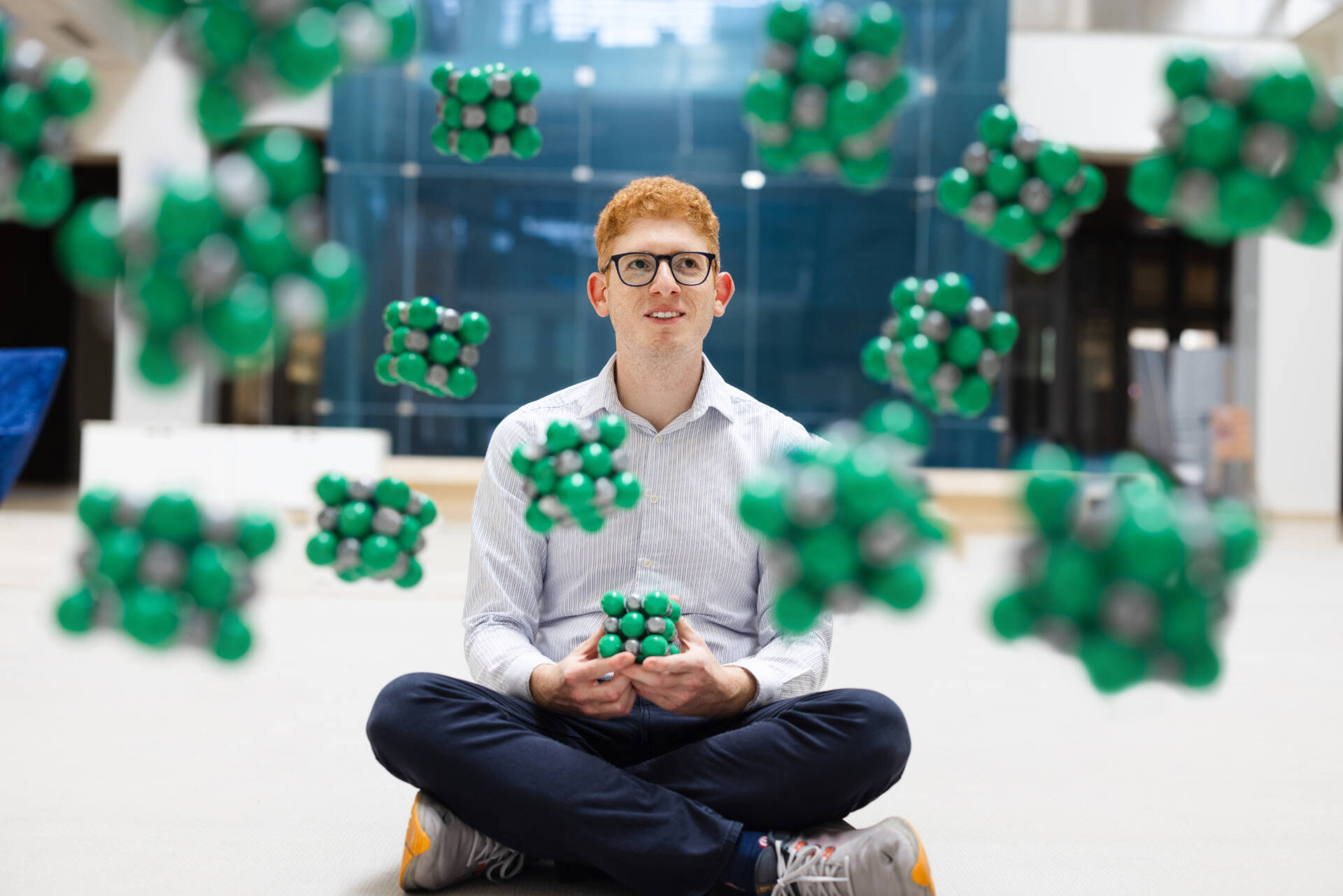
Shadi Fatayer studies matter through a microscope so powerful that individual molecules can be observed down to their component atoms, including the chemical bonds holding the structure together. The single-molecule microscopy images that he captures can look startlingly like the way organic molecules are drawn in textbooks.
“By using a scanning probe microscope, I can look at the individual atoms that constitute matter,” says Fatayer, a fundamental physics researcher who joined KAUST in 2022. Fatayer can use his microscope not only to see individual molecules, but also to interact with them, for example, triggering chemical reactions on demand at single-molecule scale.
Observing the world this way is a long-held ambition for Fatayer. “I always wanted to do high-resolution microscopy, not just for the science, but for the beauty of it,” Fatayer says. “To actually look at these systems down to the individual atoms, is one of the limits for humans,” he says.
“I always wanted to do high-resolution microscopy, not just for the science, but for the beauty of it.”
Fatayer’s ability to study materials in such detail also has highly practical applications — for example, helping to guide the work of researchers from the KAUST Solar Center to develop high-performance solar cells.
Capturing tiny beauty
Fatayer joined KAUST from IBM, the company where scanning probe microscopy was invented, and where two iconic early atomic-scale images were created. “I remember when I was eight years old, I saw these two pictures – one showed ‘IBM’ spelled out with xenon atoms and the other was a ‘quantum corral’,” Fatayer recalls.
The quantum corral, created by constructing a ring of iron atoms on a metal surface, reveals the wave-like nature of the electrons trapped within the ring. “In the circle — created by a human — you can see the standing wave, the ripples of the electrons locked inside,” Fatayer says. “And I thought, it doesn’t get more beautiful than this.”
From early work using metal atoms, the IBM Research team has more recently focused on observing and manipulating carbon-based molecules on conductive metal surfaces. “I specialize in an additional thing, to look at molecules on top of nonconductive surfaces,” Fatayer says. “It’s challenging,” he adds.
Physicists have become very good at studying systems on top of conductive surfaces, Fatayer explains. “But reality is not like this. If you look around you, most surfaces are nonconductive,” he says. For example, the catalyst particles used in arguably the most important industrial chemical reaction, the Haber-Bosch process for making synthetic fertilizer, have a nonconductive nature. To study chemical reactions at the atomic scale in a more realistic setting; we should analyze them on nonconductive surfaces, he says.
The key instrument Fatayer uses is a specialized low-temperature scanning probe microscope. “We do low-temperature microscopy because we want the molecules to be frozen in place,” Fatayer says. “A frozen molecule can give a sharper image, but there are other advantages,” he adds. Setting up a sample on a scanning microscope is a long process. “You can spend your whole day preparing your sample,” he says. But when working at room temperature, taking a break at this point is not an option because the sample will degrade with time. “Even though you are tired, you have to stay on the machine and measure, measure, measure for as long as it takes. It doesn’t allow you time to think,” he says.
Setting up a sample in a low-temperature scanning probe microscope is a more technically demanding process, Fatayer says. “But the moment it happens, that’s it. I can stop. I can think. If I am tired, I can go home,” he says. The molecule will still be frozen in position in the morning and the sample pristine. “To carry out impactful research, the most important thing is that you stop and think. This type of experiment allows you to do this. I think this is brilliant.”
“To carry out impactful research, the most important thing is that you stop and think. This type of experiment allows you to do this.”
Scanning the surface
Fatayer also uses scanning probe microscopy and X-ray photoelectron spectroscopy in collaborative projects with the KAUST Solar Center. “With this technique, you can tell how the molecules are arranged on the surface,” Fatayer says. “This is valuable information for applications such as solar cell development.”
Solar cells comprise multiple thin layers of material. “I try to help the researchers to understand how the molecules in these layers are organized,” Fatayer says. “You may have a solar cell where you make a small change to a molecule in one layer, and then suddenly the device doesn’t work,” he says. “Correlating the function of the solar cell with the molecular structure you used is something we can help with.”
The low-temperature microscopes Fatayer uses for his own atomic-scale research are not off-the-shelf items. His new instrument is due to be delivered and installed at KAUST in early 2024. Until then, Fatayer continues his work using microscopes at a small handful of collaborator institutions around the world.
One recent study, for which the microscopy work was done at IBM before Fatayer completed the theoretical component at KAUST, showed how the microscope tip could pluck the chlorine atoms from a carbon-based organic molecule to selectively control the interconversion between three different products. The study, which might inspire the development of molecular machines, is a significant scientific achievement, but for Fatayer, this is not the only reward. “For me, this work still gives me a strong artistic satisfaction,” he says.

